this is desktop
this is laptop
Ophthalmology and eye surgery in Shiraz hold a distinguished reputation in Iran and the Middle East. The city has gained widespread recognition due to the exceptional expertise of its ophthalmologists, notably led by the late Professor Ali Asghar Khodadoust. Over the years, these ophthalmologists have emerged as some of the finest in Iran and globally, delivering comprehensive diagnostic, therapeutic, and surgical services across various sub-specialties to patients from around the world, particularly in the Middle East. Notably, their expertise extends to areas such as retinal and cornea transplantation, eye correction, cataract surgery, glaucoma procedures, and more.

In the land of blinds, one eyed-man is king.
All eye services in one place.
Glaucoma is a condition affecting the eyes as one ages, characterized by the accumulation of fluid in the front part of the eye, leading to increased pressure and progressive harm to the optic nerve. Initial treatment typically involves medication and laser procedures; however, if these prove ineffective, the recommended course of action is trabeculectomy surgery. This eye surgery intervention aims to substantially reduce intraocular fluid, minimizing optic nerve damage. It achieves this by establishing a new pathway for the fluid to exit the eye more efficiently, facilitating its reabsorption by the surrounding eye tissues.
Trabeculectomy is an outpatient surgery in a specialized eye center, with the eye numbed, and the patient possibly awake. The surgeon lowers eye pressure by draining excess fluid, typically lasting one hour. Patients can return home on the same day, requiring assistance for transportation.
Trabeculectomy has a long-term success rate of approximately 90%, with two-thirds of patients no longer needing post-surgery medication to control the condition.
Recovery time varies, but most patients heal within 3 to 6 weeks. Patients can resume light job duties within one or two weeks if eye pressure normalizes. Daily activities such as reading and using electronic devices can be resumed shortly after surgery.
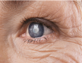
Clear and healthy vision relies on a cornea that is smooth and unimpaired. Conditions such as scarring, swelling, or damage to the cornea can disrupt the proper focusing of light into the eye, resulting in blurred vision. If attempts to heal or repair the cornea prove unsuccessful, an ophthalmologist may suggest a corneal transplant. During this procedure, the damaged cornea is substituted with a clear and healthy cornea obtained from a human donor. Corneal transplants as an eye surgery, can take various forms, including replacing only the front and middle layers, solely the inner layer, or, in certain instances, the entire cornea.
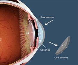
Corneal transplant procedures can use local or general anesthesia, and patients may return home on the same or next day. During the procedure, a circular piece of the damaged cornea is removed from the center and replaced with the donated cornea.
Corneal transplants are highly common, with overall success rates exceeding 90% after one year and 74% at five years.
Initial recovery spans one to three weeks, during which careful post-transplant eye care is crucial, as advised by the surgeon. Vision restoration time varies, ranging from weeks to months based on the underlying disease and individual body response.
In typical circumstances, six muscles collaborate to coordinate the alignment of both eyes. However, individuals with strabismus encounter difficulties with these muscles, causing an inability to maintain a normal eye position. Strabismus surgery involves altering the length or positioning of the eye muscles, ensuring that the eyes align correctly. This procedure aims to restore optimal vision and depth perception.
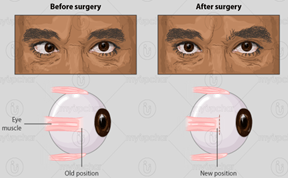
Strabismus surgery is typically an outpatient procedure lasting between 30 minutes and two hours. A membrane covering the eye muscles is opened, and each muscle is adjusted to correct vision misalignment. Patients usually need about an hour to wake up from anesthesia.
Success rates vary by age, decreasing from 85-88% in those under 50 years to 73% in adults over 50 years. For children, the surgical success rate is around 86%, and some may require a second surgery for optimal outcomes.
Recovery time depends on the procedure’s extent. Most patients can resume normal activities within a day or two, experiencing minor discomfort, bruising, and swelling. Full recovery, including eye alignment changes, typically takes six weeks.
A chalazion is a reddish lump that gradually develops on the eyelids due to the blockage of an oil gland. Usually, these lumps are swollen without a genuine infection, although occasional cases may become infected. While many chalazia resolve with the application of warm compresses, some may necessitate medical treatment or surgical drainage. The surgical procedure involves making a small incision in the lump, through which the doctor drains the fluid and eliminates the accumulated material within the nodule. Typically, stitches are not needed for this process.

Chalazion surgery is a brief office procedure taking 15 to 20 minutes. The doctor injects a numbing agent, makes a small incision in the bump, drains fluid, and removes material without stitches.
Chalazion surgery is highly effective, with a success rate exceeding 85%.
Recovery is relatively short, with the surgical incision healing in 7 to 10 days. Bruising or swelling may persist for up to two weeks, requiring caution to prevent eye injury. Resolution of the bump and redness can take up to a month.
Oculoplastic surgery is undertaken to modify the region within and around the eye, commonly employed to rectify or address a medical issue or injury, or purely for cosmetic reasons. This encompasses surgical procedures involving the eyelids, the bony structures surrounding the eye, and the tear duct system.

The use of a prosthetic eye can enhance the aesthetic appearance of individuals who have suffered eye loss due to injuries or illnesses like cancer, infections, or glaucoma. In cases where surgical removal of an eye is necessary, implanting a prosthetic eye is typically advised. This implant aids in maintaining the normal functioning of the eyelid. During the procedure, the doctor connects the muscles that used to control the natural eye to a permanently embedded ocular implant in the eye socket. This ocular implant serves as support for the prosthetic eye placed over it.

The optic nerve and eye muscles are cut, the eyeball is replaced with an implant shaped like a ball, and the muscles are re-attached to the eye implant. After healing, a prosthetic eye can be fitted.
Approximately 98% of patients with an intraocular prosthesis have a successful outcome.
Healing takes six to eight weeks for most patients, with a prosthetic eye fitted after recovery.
Retinal detachment is a critical condition wherein the thin tissue layer, the retina, detaches from its usual position. This separation results in a lack of oxygen and nutrients for the retinal cells from the blood vessels, potentially leading to vision loss in the affected eye if left untreated. Surgical intervention is typically the essential solution to address retinal tears, holes, or detachments. Surgical techniques employed in these cases include suturing a piece of silicone material over the affected area, injecting air or gas to press the hole against the eye wall, and draining and replacing the fluid in the eye.
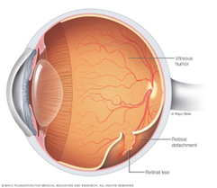
The chosen surgical technique depends on the severity of detachment. Suturing silicone material, injecting air or gas, and draining and replacing eye fluid are common techniques.
Retinal detachment surgery has a success rate of approximately 90%, with some cases requiring more than one operation.
Vision recovery occurs over several weeks or months following surgery, and full recovery is not immediate. Time is needed for the retina to recover.
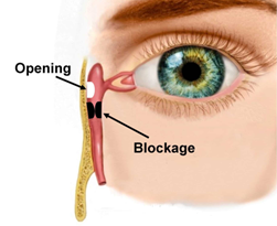
DCR surgery can be external or endonasal, typically taking 30 minutes to two hours. Endonasal DCR has a higher success rate (90-98%) compared to external DCR (83-85%).
Most patients can go home the same day, with mild bleeding and fatigue lasting 1-2 days and 1-2 weeks, respectively. Postoperative symptoms, such as a bloody discharge, usually subside within a few days. Heavy lifting should be avoided for 7-10 days.

Laser photocoagulation, delivering intense light energy, takes minutes, and patients can return home afterward.
Laser photocoagulation reduces severe visual loss risk by 60-73% in two years, with additional sessions based on the condition’s severity.
Full recovery takes weeks, with the need for sunglasses due to dilated eyes. Stable blood glucose levels are crucial for long-term symptom control.
Proptosis refers to the protrusion of one or both eyes beyond their normal position, resulting from an augmentation in the contents of the orbital space within the regular bony orbit anatomy, causing forward displacement of the eye. Bulging eyes, often associated with thyroid issues, can be addressed with oral and intravenous medications. Surgical intervention is also utilized for managing proptosis, involving the removal of the problematic tissue responsible for this condition, along with the repair of ocular muscles.

Endoscopic orbital decompression is performed through the nostrils, while lateral orbital decompression requires cutting the skin.
Recovery takes 7-10 days, with prescription pain medication to manage discomfort during the initial days.
A pterygium is a benign growth of the thin mucous membrane extending beyond the cornea. Treatment may not be necessary if the lesion is asymptomatic. However, if it starts causing vision issues impacting daily activities, removal of the pterygium may be required. During pterygium surgery, the lesion is extracted from the eye’s surface, and a small piece of skin from beneath the eyelid is used to fill the area from which the pterygium was removed..

Pterygium surgery lasts 30-45 minutes, and different techniques have varying recurrence rates.
Recovery can take weeks to months, with an eye patch applied after surgery to prevent infection and discomfort.

CXL involves applying vitamin B2 and controlled ultraviolet light to eliminate corneal ectasia.
Studies report a 99% stability or improvement in corneal shape with both CXL types.
After Epi-off CXL, eye soreness lasts 5-7 days, requiring a week off work. Continued surface discomfort is common in the early months after most eye surgeries.

HAPPY GREEN LIFE Co. ©2023 All rights reserved
FREE CONSULTATION
FREE CONSULTATION
Hi! Click one of our members below to chat on WhatsApp:
Our consulting team will send you a message after reviewing your request